Your F actin staining images are ready. F actin staining are a topic that is being searched for and liked by netizens today. You can Download the F actin staining files here. Get all royalty-free photos.
If you’re looking for f actin staining images information related to the f actin staining topic, you have pay a visit to the right site. Our site always gives you hints for seeing the maximum quality video and image content, please kindly hunt and locate more enlightening video articles and graphics that fit your interests.
F Actin Staining. Here we describe Lifeact a 17-amino-acid peptide which stained filamentous actin F-actin structures in eukaryotic cells an. Actin is one of the most abundant proteins found in cells and can be labeled with a fluorophore very easily when the monomers form. For successful phalloidin staining F-actin must be fixed and maintained in its native conformation. For the observation of F-actin-related processes non-invasive live cell imaging has become the state-of-the-art technique.
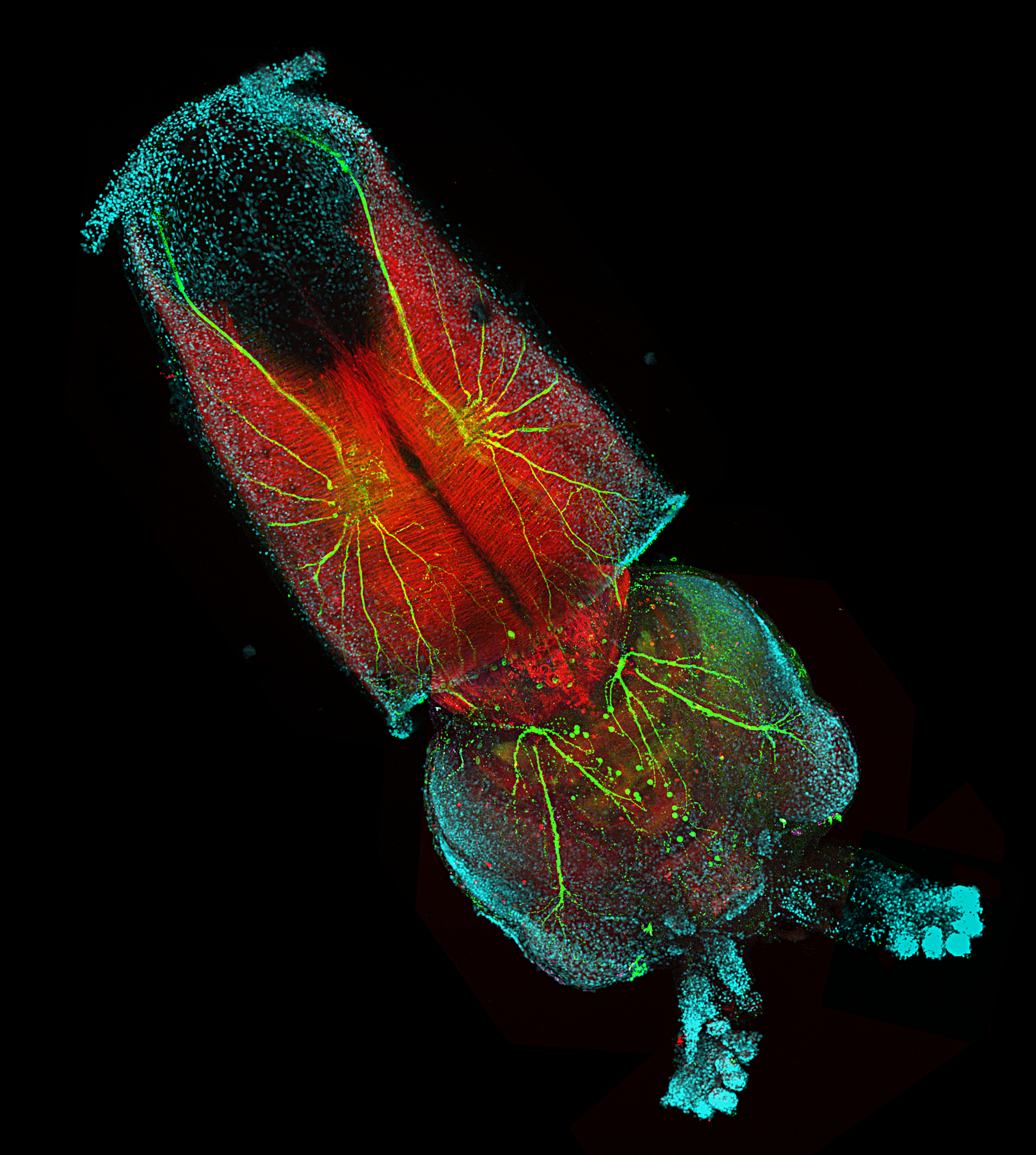
 Longfin Inshore Squid From pinterest.com
Paraformaldehyde or some derivative usually at 4 concentration is an excellent fixative for this purpose. The Factin staining pattern in osteoclasts treated with cytochalasin B appeared as a small punctate or diffuse pattern throughout the cytoplasm. Here we describe Lifeact a 17-amino-acid peptide which stained filamentous actin F-actin structures in eukaryotic cells an. As compared with the staining pattern of Factin Gactin appeared as a more densely stained perinuclear areas. Actin is one of the most abundant proteins found in cells and can be labeled with a fluorophore very easily when the monomers form. This is a video teaching aid developed to supplement the text protocolThis is Part 2 of BS1007 Cytochemistry.
Invitrogen CellMask Deep Red Actin Tracking Stain fluorescently labels polymerizedfilamentous actin F-actin in live or fixed cells.
For the observation of F-actin-related processes non-invasive live cell imaging has become the state-of-the-art technique. As compared with the staining pattern of Factin Gactin appeared as a more densely stained perinuclear areas. Paraformaldehyde or some derivative usually at 4 concentration is an excellent fixative for this purpose. The selective staining of cell compartments provides a powerful method for studying cellular events in a spatial and temporal. The Factin staining pattern in osteoclasts treated with cytochalasin B appeared as a small punctate or diffuse pattern throughout the cytoplasm. Invitrogen CellMask Deep Red Actin Tracking Stain fluorescently labels polymerizedfilamentous actin F-actin in live or fixed cells.
 Source: pinterest.com
In the cytoD treated hMSCs F-actin intensity was higher on the cell border than in the cell center due to a lack of stress fibers. As compared with the staining pattern of Factin Gactin appeared as a more densely stained perinuclear areas. In the untreated hMSCs actin was distributed equally over the cell. Rhodamine-phalloidin was used to label F-actin in unfixed cells of 13 species of filamentous and blade-forming red algae from the three families Ceramiaceae Acrochaetiaceae and Bangiaceae. Paraformaldehyde or some derivative usually at 4 concentration is an excellent fixative for this purpose.
 Source: pinterest.com
Immunofluorescent staining has been most frequently used to study cytoskeletal components. Fluorescent and biotinylated phallotoxins stain F-actin at nanomolar concentrations and are extremely water soluble thus providing convenient probes for labeling identifying and quantitating F-actin in tissue sections cell cultures or cell-free experiments. The selective staining of cell compartments provides a powerful method for studying cellular events in a spatial and temporal. Methanol is not a good choice for phalloidin staining because F-actins conformation is lost with that fixative. Typically it is used conjugated to a fluorescent dye such as FITC Rhodamine TRITC or similar dyes such as Alexa Fluor 488 or iFluor 488.
 Source: pinterest.com
It binds to all variants of actin filaments in many different species of animals and plants. Rhodamine-phalloidin was used to label F-actin in unfixed cells of 13 species of filamentous and blade-forming red algae from the three families Ceramiaceae Acrochaetiaceae and Bangiaceae. F-actin visualization using fluorescent markers is an important tool for getting a deeper understanding of the structural cytoskeletal dynamics. For successful phalloidin staining F-actin must be fixed and maintained in its native conformation. CellMask Deep Red Actin Tracking Stain can be used to track F-actin in live cells for 24 hours or more.
 Source: pinterest.com
The Factin staining pattern in osteoclasts treated with cytochalasin B appeared as a small punctate or diffuse pattern throughout the cytoplasm. For successful phalloidin staining F-actin must be fixed and maintained in its native conformation. Paraformaldehyde or some derivative usually at 4 concentration is an excellent fixative for this purpose. View All 398 Related Papers 1. Actin is one of the most abundant proteins found in cells and can be labeled with a fluorophore very easily when the monomers form.
 Source: pinterest.com
The selective staining of cell compartments provides a powerful method for studying cellular events in a spatial and temporal. As expected the distribution of F-actin changed upon treatment with cytoD Fig. Fluorescent and biotinylated phallotoxins stain F-actin at nanomolar concentrations and are extremely water soluble thus providing convenient probes for labeling identifying and quantitating F-actin in tissue sections cell cultures or cell-free experiments. The cytoskeleton is composed of a series of filamentous structures including intermediate filaments actin filaments and microtubules. In the cytoD treated hMSCs F-actin intensity was higher on the cell border than in the cell center due to a lack of stress fibers.
 Source: in.pinterest.com
In the cytoD treated hMSCs F-actin intensity was higher on the cell border than in the cell center due to a lack of stress fibers. Typically it is used conjugated to a fluorescent dye such as FITC Rhodamine TRITC or similar dyes such as Alexa Fluor 488 or iFluor 488. Labelling was achieved only after treatment with either beta-glucuronidase or a combination of cellulase and an extract of snail gut enzyme. It binds to all variants of actin filaments in many different species of animals and plants. Paraformaldehyde or some derivative usually at 4 concentration is an excellent fixative for this purpose.
 Source: pinterest.com
It binds to all variants of actin filaments in many different species of animals and plants. Phalloidin is a highly selective bicyclic peptide that is used for staining actin filaments also known as F-actin. The selective staining of cell compartments provides a powerful method for studying cellular events in a spatial and temporal. The fixation procedure is critical for obtaining faithful representation of the F-actin distribution within the cell. Less F-actin was found over the nucleus Fig.
 Source: pinterest.com
Therefore in cell biology the study of organization and structurefunction relationships is of great importance. Such a punctate pattern of Factin staining is almost corresponding to those of Gactin Figs. As expected the distribution of F-actin changed upon treatment with cytoD Fig. Therefore in cell biology the study of organization and structurefunction relationships is of great importance. F-actin visualization using fluorescent markers is an important tool for getting a deeper understanding of the structural cytoskeletal dynamics.
 Source: pinterest.com
This staining procedure is particularly suitable for staining the male fusome and the cytokinetic contractile ring. Paraformaldehyde or some derivative usually at 4 concentration is an excellent fixative for this purpose. Preparations of Drosophila testes fixed with paraformaldehyde can be stained for F-actin according to the protocol described here. Protocol 1 - Fixing and staining tissue culture cells with fluorescent phalloidin F-actin Dapi DNA and an antibody. 1-3 Labeled phallotoxins have similar affinity for both large and small filaments binding in a stoichiometric ratio of about one.
 Source: nl.pinterest.com
View All 398 Related Papers 1. This staining procedure is particularly suitable for staining the male fusome and the cytokinetic contractile ring. Less F-actin was found over the nucleus Fig. The cytoskeleton is composed of a series of filamentous structures including intermediate filaments actin filaments and microtubules. Protocol 1 - Fixing and staining tissue culture cells with fluorescent phalloidin F-actin Dapi DNA and an antibody.
 Source: ar.pinterest.com
Immunofluorescent staining has been most frequently used to study cytoskeletal components. The Factin staining pattern in osteoclasts treated with cytochalasin B appeared as a small punctate or diffuse pattern throughout the cytoplasm. Here we describe Lifeact a 17-amino-acid peptide which stained filamentous actin F-actin structures in eukaryotic cells an. Less F-actin was found over the nucleus Fig. Then stain ovules overnight at 4C with 033 μM rhodamine-phalloidin for labeling F-actin and 3 μgml Hoechst 33258 to counterstain cell nuclei diluted in blocking solution.
 Source: pinterest.com
Preparations of Drosophila testes fixed with paraformaldehyde can be stained for F-actin according to the protocol described here. It is designed to readily permeate live cells thus providing more uniform and bright labeling. For successful phalloidin staining F-actin must be fixed and maintained in its native conformation. The Factin staining pattern in osteoclasts treated with cytochalasin B appeared as a small punctate or diffuse pattern throughout the cytoplasm. Then stain ovules overnight at 4C with 033 μM rhodamine-phalloidin for labeling F-actin and 3 μgml Hoechst 33258 to counterstain cell nuclei diluted in blocking solution.
 Source: pinterest.com
Then stain ovules overnight at 4C with 033 μM rhodamine-phalloidin for labeling F-actin and 3 μgml Hoechst 33258 to counterstain cell nuclei diluted in blocking solution. CellMask Deep Red Actin Tracking Stain can be used to track F-actin in live cells for 24 hours or more. 1-3 Labeled phallotoxins have similar affinity for both large and small filaments binding in a stoichiometric ratio of about one. Actin is one of the most abundant proteins found in cells and can be labeled with a fluorophore very easily when the monomers form. This staining procedure is particularly suitable for staining the male fusome and the cytokinetic contractile ring.
 Source: pinterest.com
Invitrogen CellMask Deep Red Actin Tracking Stain fluorescently labels polymerizedfilamentous actin F-actin in live or fixed cells. CellMask Deep Red Actin Tracking Stain can be used to track F-actin in live cells for 24 hours or more. Paraformaldehyde or some derivative usually at 4 concentration is an excellent fixative for this purpose. It binds to all variants of actin filaments in many different species of animals and plants. Protocol 1 - Fixing and staining tissue culture cells with fluorescent phalloidin F-actin Dapi DNA and an antibody.
 Source: pinterest.com
Rhodamine-phalloidin was used to label F-actin in unfixed cells of 13 species of filamentous and blade-forming red algae from the three families Ceramiaceae Acrochaetiaceae and Bangiaceae. In the cytoD treated hMSCs F-actin intensity was higher on the cell border than in the cell center due to a lack of stress fibers. The selective staining of cell compartments provides a powerful method for studying cellular events in a spatial and temporal. Less F-actin was found over the nucleus Fig. Then stain ovules overnight at 4C with 033 μM rhodamine-phalloidin for labeling F-actin and 3 μgml Hoechst 33258 to counterstain cell nuclei diluted in blocking solution.
 Source: pinterest.com
Rhodamine-phalloidin was used to label F-actin in unfixed cells of 13 species of filamentous and blade-forming red algae from the three families Ceramiaceae Acrochaetiaceae and Bangiaceae. Dehydration After the staining period NB3 dehydrate ovules in isopropanol NB4 increasing-concentration solutions 75 85 95 and 100 for 7 min each at 4C and finally in 100 isopropanol for 1012 min also at 4C. Phalloidin is a highly selective bicyclic peptide that is used for staining actin filaments also known as F-actin. Live imaging of the actin cytoskeleton is crucial for the study of many fundamental biological processes but current approaches to visualize actin have several limitations. Labelling was achieved only after treatment with either beta-glucuronidase or a combination of cellulase and an extract of snail gut enzyme.
 Source: pinterest.com
It is designed to readily permeate live cells thus providing more uniform and bright labeling. 1-3 Labeled phallotoxins have similar affinity for both large and small filaments binding in a stoichiometric ratio of about one. The Factin staining pattern in osteoclasts treated with cytochalasin B appeared as a small punctate or diffuse pattern throughout the cytoplasm. In the untreated hMSCs actin was distributed equally over the cell. Dehydration After the staining period NB3 dehydrate ovules in isopropanol NB4 increasing-concentration solutions 75 85 95 and 100 for 7 min each at 4C and finally in 100 isopropanol for 1012 min also at 4C.
 Source: pinterest.com
Here we describe Lifeact a 17-amino-acid peptide which stained filamentous actin F-actin structures in eukaryotic cells an. 1-3 Labeled phallotoxins have similar affinity for both large and small filaments binding in a stoichiometric ratio of about one. It binds to all variants of actin filaments in many different species of animals and plants. View All 398 Related Papers 1. For successful phalloidin staining F-actin must be fixed and maintained in its native conformation.
This site is an open community for users to submit their favorite wallpapers on the internet, all images or pictures in this website are for personal wallpaper use only, it is stricly prohibited to use this wallpaper for commercial purposes, if you are the author and find this image is shared without your permission, please kindly raise a DMCA report to Us.
If you find this site adventageous, please support us by sharing this posts to your favorite social media accounts like Facebook, Instagram and so on or you can also save this blog page with the title f actin staining by using Ctrl + D for devices a laptop with a Windows operating system or Command + D for laptops with an Apple operating system. If you use a smartphone, you can also use the drawer menu of the browser you are using. Whether it’s a Windows, Mac, iOS or Android operating system, you will still be able to bookmark this website.


