Your How to time lapse of liqid droplet protein images are available in this site. How to time lapse of liqid droplet protein are a topic that is being searched for and liked by netizens today. You can Find and Download the How to time lapse of liqid droplet protein files here. Get all royalty-free photos.
If you’re searching for how to time lapse of liqid droplet protein images information linked to the how to time lapse of liqid droplet protein keyword, you have come to the right blog. Our site frequently gives you hints for refferencing the highest quality video and picture content, please kindly hunt and find more informative video articles and images that fit your interests.
How To Time Lapse Of Liqid Droplet Protein. 44 Select individual cells in the monolayer cultures for analysis. The formation of complexes could be inhibited by the nocodazole-induced depolymerization of the microtubules. Select the first dividing cell after 34 h of incubation following the start of time-lapse microscopy. D and E show typical protein aggregates in the turbid liquid containing proteins that do not undergo LLPS but form aggregates.
Localization Of The Triacylglycerol Lipase Tgl3 On A Subpopulation Of Download Scientific Diagram From researchgate.net
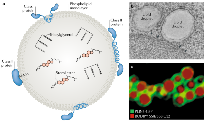
Visualization of LD biogenesis and dynamics in transfected cells. Turbidity assay together with time-lapse imaging of liquid droplet formation to monitor cGAMP production and multivalency-induced liquid-phase condensation in this system. Long time-lapse recording periods longer than 24 h require that the embryo be mounted in a stable setting such as in agarose. Time-lapse melting of benzoic acid. And 3 droplets formed by a pathogenic mutant of hnRNPA1. Allow the cells adaptation time after starting time-lapse microscopy.
The values of A were obtained using bcHdef and averaged over 4 nanoclusters.
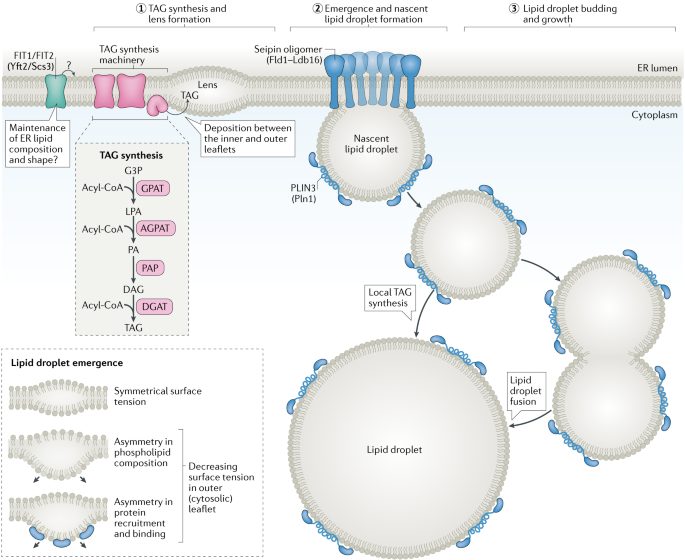
Thus the process is dependent on microtubules. To prevent the agarose from floating off the coverslip during long imaging periods a plastic mesh can be glued onto the coverslip with silicon grease before the application of the embryo within the liquid agarose. After centrifuging at 17000 g for 20 min at 4 C the pellet was resuspended in 15 ml of CSF-XB with 10 sucrose wv 10 μg each of leupeptin pepstatin and chymostatin 1 mM GTP 1 mM DTT. A scaling analysis indicates that nanostar liquids that are farther from the phase boundary ie farther from the dissolution or boiling temperature T b are degraded only on the droplet surface while those poised near the boundary permit enzyme penetration. Lipid droplets can grow after they have been assembled. The presence of dynein on ADRP-containing droplets supports a role for this motor protein.
Source: researchgate.net
Allow the cells adaptation time after starting time-lapse microscopy. Visualization of LD biogenesis and dynamics in transfected cells. In biology intracellular LLPS is a phenomenon in which highly concentrated liquid phases of certain proteins or biomolecules are generated as liquid droplets inside a. In starved cells GFPADRP was homogenously dispersed in the cell cytosol and nucleus and it was occasionally detected in the plasma membrane see Figure 6C. To prevent the agarose from floating off the coverslip during long imaging periods a plastic mesh can be glued onto the coverslip with silicon grease before the application of the embryo within the liquid agarose.
 Source: nature.com
Since the Ninth Drop fell in April 2014 the Tenth Drop has start. Thus the process is dependent on microtubules. Since the Ninth Drop fell in April 2014 the Tenth Drop has start. We suggest that proximity to the phase boundary accelerates liquid. Supplementary Figure 8 Time-lapse micrographs obtained by in situ liquid TEM of HBP-2 protein at a salt concentration NaCl 0137 M and with increasing electron dose rate indicated on the right showing an increased growth rate A of protein nanoclusters.
 Source: europepmc.org
The formation of complexes could be inhibited by the nocodazole-induced depolymerization of the microtubules. We have also investigated theimpact of cGAS mut ations of basicresidueslocated on the cGASDNA site-C interface including a pair of tumor. F Time-lapse analysis showing that phase-separated liquid droplets formed by 3 μmolL SEPA-1PGL-1-3 fuse with each other red arrowhead and relax into a larger one. Time-lapse melting of benzoic acid. In this time-lapse composite image a water droplet is propelled into the air by an oscillating superhydrophobic surface.
Source: researchgate.net
1d n 6 independent experiments. The worlds longest running experiment the Pitch Drop - Time lapse April 2012 - April 2015. The time lapse VCR had the capability of compressing real time events from 1 to 240-fold so that maximally 240 h could be recorded on a single 2 h magnetic tape. 44 Select individual cells in the monolayer cultures for analysis. Time-lapse movie stills from the first 15 min after irradiation are shown.
 Source: nature.com
Visualization of LD biogenesis and dynamics in transfected cells. After centrifuging at 17000 g for 20 min at 4 C the pellet was resuspended in 15 ml of CSF-XB with 10 sucrose wv 10 μg each of leupeptin pepstatin and chymostatin 1 mM GTP 1 mM DTT. Visualization of LD biogenesis and dynamics in transfected cells. White arrows indicate the orientation of the laser line. In general we found that a compression of 72-fold was optimal for our experiments.
 Source: nature.com
Select the first dividing cell after 34 h of incubation following the start of time-lapse microscopy. If the droplets internal vibration has a frequency about three times that of the surfaces rise and fall its initial kinetic energy can be boosted by as much as 250. Ex1 proteins with different polyQlengths2543or97fusedtoaC-terminaleGFPtagFig-ure 1A in HEK293 cells and followed their expression by time-lapse fluorescence microscopy for 2448 hr. In biology intracellular LLPS is a phenomenon in which highly concentrated liquid phases of certain proteins or biomolecules are generated as liquid droplets inside a. Time-lapse movie stills from the first 15 min after irradiation are shown.
 Source: cell.com
We will refer to these proteins as 25 43 or 97QP-GFP where the number indi-cates the polyQ length eg 97Q and the P indicates the C-ter-. After centrifuging at 17000 g for 20 min at 4 C the pellet was resuspended in 15 ml of CSF-XB with 10 sucrose wv 10 μg each of leupeptin pepstatin and chymostatin 1 mM GTP 1 mM DTT. The worlds longest running experiment the Pitch Drop - Time lapse April 2012 - April 2015. White arrows indicate the orientation of the laser line. In this time-lapse composite image a water droplet is propelled into the air by an oscillating superhydrophobic surface.
 Source: europepmc.org
If the droplets internal vibration has a frequency about three times that of the surfaces rise and fall its initial kinetic energy can be boosted by as much as 250. And 3 droplets formed by a pathogenic mutant of hnRNPA1. Time-lapse melting of benzoic acid. In general we found that a compression of 72-fold was optimal for our experiments. Lipid droplets can grow after they have been assembled.
 Source: researchgate.net
Flying extra high. Ex1 proteins with different polyQlengths2543or97fusedtoaC-terminaleGFPtagFig-ure 1A in HEK293 cells and followed their expression by time-lapse fluorescence microscopy for 2448 hr. Lipid droplets can grow after they have been assembled. Visualization of LD biogenesis and dynamics in transfected cells. The worlds longest running experiment the Pitch Drop - Time lapse April 2012 - April 2015.
 Source: jbc.org
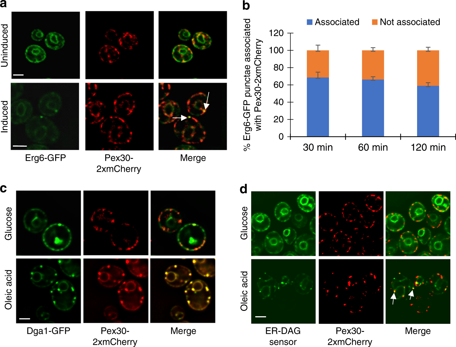
The worlds longest running experiment the Pitch Drop - Time lapse April 2012 - April 2015. A Time-lapse analysis of COS7 cells transfected with an inactive form of GFP tagged TBC1D20 as an ER membrane marker green and upper mid-panel ER treated with OA and labeled with BODIPY red and lower mid panel using a peristaltic pump as described in Fig. 44 Select individual cells in the monolayer cultures for analysis. Maturation appears to be driven at least in part by the formation of amyloid-like fibers because 1 over time many of the IDR proteins form filaments that can be observed by light andor electron microscopy Figures 2C and 3B. Select the first dividing cell after 34 h of incubation following the start of time-lapse microscopy.
 Source: researchgate.net
Ex1 proteins with different polyQlengths2543or97fusedtoaC-terminaleGFPtagFig-ure 1A in HEK293 cells and followed their expression by time-lapse fluorescence microscopy for 2448 hr. Flying extra high. Select the first dividing cell after 34 h of incubation following the start of time-lapse microscopy. Maturation appears to be driven at least in part by the formation of amyloid-like fibers because 1 over time many of the IDR proteins form filaments that can be observed by light andor electron microscopy Figures 2C and 3B. 44 Select individual cells in the monolayer cultures for analysis.
 Source: europepmc.org
Select the first dividing cell after 34 h of incubation following the start of time-lapse microscopy. Turbidity assay together with time-lapse imaging of liquid droplet formation to monitor cGAMP production and multivalency-induced liquid-phase condensation in this system. 44 Select individual cells in the monolayer cultures for analysis. Long time-lapse recording periods longer than 24 h require that the embryo be mounted in a stable setting such as in agarose. We suggest that proximity to the phase boundary accelerates liquid.
 Source: cell.com
Allow the cells adaptation time after starting time-lapse microscopy. We find that the degradation rate strongly changes as a function of the collective stability of the liquid phase. For observation of liquid droplet behaviours z-stack time-lapses were taken for 510 min. The worlds longest running experiment the Pitch Drop - Time lapse April 2012 - April 2015. The formation of complexes could be inhibited by the nocodazole-induced depolymerization of the microtubules.
 Source: jbc.org
Flying extra high. Turbidity assay together with time-lapse imaging of liquid droplet formation to monitor cGAMP production and multivalency-induced liquid-phase condensation in this system. Introduction Various physiological processes are currently being explored using liquidliquid phase separation LLPS. White arrows indicate the orientation of the laser line. After centrifuging at 17000 g for 20 min at 4 C the pellet was resuspended in 15 ml of CSF-XB with 10 sucrose wv 10 μg each of leupeptin pepstatin and chymostatin 1 mM GTP 1 mM DTT.
 Source: researchgate.net
White arrows indicate the orientation of the laser line. Allow the cells adaptation time after starting time-lapse microscopy. The worlds longest running experiment the Pitch Drop - Time lapse April 2012 - April 2015. 12 LLPS is a phenomenon in which two or more different mixtures are separated into multiple liquid phases. To prevent the agarose from floating off the coverslip during long imaging periods a plastic mesh can be glued onto the coverslip with silicon grease before the application of the embryo within the liquid agarose.
 Source: mdpi.com
Turbidity assay together with time-lapse imaging of liquid droplet formation to monitor cGAMP production and multivalency-induced liquid-phase condensation in this system. Long time-lapse recording periods longer than 24 h require that the embryo be mounted in a stable setting such as in agarose. In general we found that a compression of 72-fold was optimal for our experiments. 1d n 6 independent experiments. Thus the process is dependent on microtubules.
 Source: researchgate.net
To prevent the agarose from floating off the coverslip during long imaging periods a plastic mesh can be glued onto the coverslip with silicon grease before the application of the embryo within the liquid agarose. Long time-lapse recording periods longer than 24 h require that the embryo be mounted in a stable setting such as in agarose. The presence of dynein on ADRP-containing droplets supports a role for this motor protein. After centrifuging at 17000 g for 20 min at 4 C the pellet was resuspended in 15 ml of CSF-XB with 10 sucrose wv 10 μg each of leupeptin pepstatin and chymostatin 1 mM GTP 1 mM DTT. Allow the cells adaptation time after starting time-lapse microscopy.
Source: researchgate.net
If the droplets internal vibration has a frequency about three times that of the surfaces rise and fall its initial kinetic energy can be boosted by as much as 250. Turbidity assay together with time-lapse imaging of liquid droplet formation to monitor cGAMP production and multivalency-induced liquid-phase condensation in this system. To prevent the agarose from floating off the coverslip during long imaging periods a plastic mesh can be glued onto the coverslip with silicon grease before the application of the embryo within the liquid agarose. In general we found that a compression of 72-fold was optimal for our experiments. After centrifuging at 17000 g for 20 min at 4 C the pellet was resuspended in 15 ml of CSF-XB with 10 sucrose wv 10 μg each of leupeptin pepstatin and chymostatin 1 mM GTP 1 mM DTT.
This site is an open community for users to do sharing their favorite wallpapers on the internet, all images or pictures in this website are for personal wallpaper use only, it is stricly prohibited to use this wallpaper for commercial purposes, if you are the author and find this image is shared without your permission, please kindly raise a DMCA report to Us.
If you find this site serviceableness, please support us by sharing this posts to your preference social media accounts like Facebook, Instagram and so on or you can also bookmark this blog page with the title how to time lapse of liqid droplet protein by using Ctrl + D for devices a laptop with a Windows operating system or Command + D for laptops with an Apple operating system. If you use a smartphone, you can also use the drawer menu of the browser you are using. Whether it’s a Windows, Mac, iOS or Android operating system, you will still be able to bookmark this website.


