Your Protein diffusion coefficient images are ready. Protein diffusion coefficient are a topic that is being searched for and liked by netizens today. You can Get the Protein diffusion coefficient files here. Get all royalty-free photos and vectors.
If you’re looking for protein diffusion coefficient pictures information related to the protein diffusion coefficient topic, you have visit the ideal site. Our site frequently gives you suggestions for seeking the highest quality video and picture content, please kindly hunt and find more enlightening video articles and graphics that match your interests.
Protein Diffusion Coefficient. Out the protein of interest and simultaneously provide its diffusion coefficient in terms of that proteins measured retention time. The electrode area can be determined electrochemically with equations equivalent to an equation and by using a redox couple having a known diffusion coefficient. A 4-l 4Cl labelled lipoprotein. No calibration is necessary.
Https Www Cell Com Biophysj Pdfextended S0006 3495 19 30048 7 From
These results are compared to dynamic light scattering approaches which are limited to k D determinations for solutions of pure protein. Those associated with the earlier data have been inferred from their apparent reproducibility. Molecular translational self-diffusion a measure of diffusive motion provides information on the effective molecular hydrodynamic radius as well as information on the properties of media or solution through which the molecule. MW 25 kDa pI 875. For biological molecules the diffusion coefficients normally range from 10 11 to 10 10 m 2 s. A simple apparatus was used to measure the diffusion coefficients of several small solutes and 18 proteins.
No calibration is necessary.
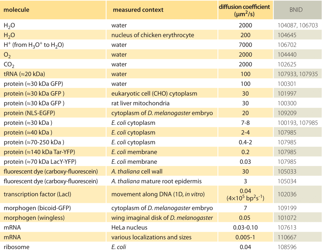
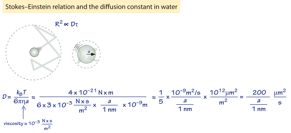
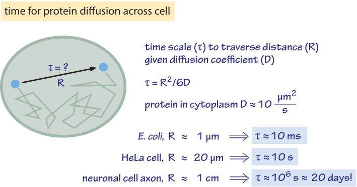
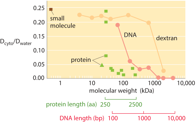
As derived in Figure 1 the characteristic diffusion constant for a molecule the size of a monomeric protein is 100 µm 2 s in water and is about ten-fold smaller 10 µm 2 s inside a cell with large variations depending on the cellular context as shown in Table 1 larger proteins often show another order of magnitude decrease to 1 µm 2 s BNID 107985. 214 g100 ml PH 74. The diffusion coefficient is the proportionality between flux and concentration gradient. 100 mglOO ml PH 74. Diffusion coefficient of plasma membrane protein. This method can also explore cross-term diffusion coefficient effects.
 Source: sciencedirect.com
A simple apparatus was used to measure the diffusion coefficients of several small solutes and 18 proteins. As derived in Figure 1 the characteristic diffusion constant for a molecule the size of a monomeric protein is 100 µm 2 s in water and is about ten-fold smaller 10 µm 2 s inside a cell with large variations depending on the cellular context as shown in Table 1 larger proteins often show another order of magnitude decrease to 1 µm 2 s BNID 107985. Diffusion Coefficient Protein Software Convective Adsorption - Desorption v21 The software is used to study water diffusion processes with known experimental data for the following geometries. Diffusion Coefficient n Many drugs have a low molecular wt q 100 MW 500 gmol q Not true for proteins and larger molecules n Examples Insulin 41000 83 x 10-7 Huge size Caffeine 1942 49 x 10-6 Medium size Dcm2s Note Aqueous Drug MW gmol. Effective diffusion coefficients of a therapeutic protein of interest BMP-2 were predicted for each hydrogel using the diffusion coefficient of αCT which has a similar molecular weight and isoelectric point to BMP-2 αCT.
 Source: book.bionumbers.org
Diffusion Coefficient n Many drugs have a low molecular wt q 100 MW 500 gmol q Not true for proteins and larger molecules n Examples Insulin 41000 83 x 10-7 Huge size Caffeine 1942 49 x 10-6 Medium size Dcm2s Note Aqueous Drug MW gmol. MW 25 kDa pI 875. The diffusion coefficient of lysozyme a globular protein was measured at various conditions as functions of lysozyme concentration salt concentration and solution age in concentrated saturated and supersaturated solutions employing Gouy interferometry. Diffusion coefficient of microRNA in cell. Each determination required 7-90 min depending on the magnitude of the diffusion coefficient.
 Source: book.bionumbers.org
Each determination required 7-90 min depending on the magnitude of the diffusion coefficient. As derived in Figure 1 the characteristic diffusion constant for a molecule the size of a monomeric protein is 100 µm 2 s in water and is about ten-fold smaller 10 µm 2 s inside a cell with large variations depending on the cellular context as shown in Table 1 larger proteins often show another order of magnitude decrease to 1 µm 2 s BNID 107985. Most results were within - 10 of literature values. These results are compared to dynamic light scattering approaches which are limited to k D determinations for solutions of pure protein. At higher protein-to lipid ratios up to 3000 μm 2 2 and use of diffusion measurements for protein geometry size oligomerization.
 Source: slidetodoc.com
105180 Diffusion coefficient of oxygen in water. The electrode area can be determined electrochemically with equations equivalent to an equation and by using a redox couple having a known diffusion coefficient. 114984 Diffusion coefficient of glycerol in water at 25C. Measuring translational diffusion coefficients of peptides and proteins by PFG-NMR using band-selective RF pulses. 105180 Diffusion coefficient of oxygen in water.
 Source: sciencedirect.com
114984 Diffusion coefficient of glycerol in water at 25C. We characterize protein diffusion by photobleaching whole cells at a single point and imaging the concentration change of fluorescent-labeled protein throughout the cell as a. Diffusion Coefficient Protein Software Convective Adsorption - Desorption v21 The software is used to study water diffusion processes with known experimental data for the following geometries. MW 25 kDa pI 875. The self-diffusion coefficient of protein molecules in solution as a function of temperature.
 Source: sciencedirect.com
As derived in Figure 1 the characteristic diffusion constant for a molecule the size of a monomeric protein is 100 µm 2 s in water and is about ten-fold smaller 10 µm 2 s inside a cell with large variations depending on the cellular context as shown in Table 1 larger proteins often show another order of magnitude decrease to 1 µm 2 s BNID 107985. Diffusion coefficient of GFP. Effective diffusion coefficients of a therapeutic protein of interest BMP-2 were predicted for each hydrogel using the diffusion coefficient of αCT which has a similar molecular weight and isoelectric point to BMP-2 αCT. 100 mglOO ml PH 74. 214 g100 ml PH 74.
 Source: pubs.rsc.org
The diffusion coefficient of lysozyme a globular protein was measured at various conditions as functions of lysozyme concentration salt concentration and solution age in concentrated saturated and supersaturated solutions employing Gouy interferometry. Measuring translational diffusion coefficients of peptides and proteins by PFG-NMR using band-selective RF pulses. The software makes possible the determination of the. Most results were within - 10 of literature values. Effective diffusion coefficients of a therapeutic protein of interest BMP-2 were predicted for each hydrogel using the diffusion coefficient of αCT which has a similar molecular weight and isoelectric point to BMP-2 αCT.
 Source: researchgate.net
Those associated with the earlier data have been inferred from their apparent reproducibility. Diffusion coefficient of GFP. At higher protein-to lipid ratios up to 3000 μm 2 2 and use of diffusion measurements for protein geometry size oligomerization. These results are compared to dynamic light scattering approaches which are limited to k D determinations for solutions of pure protein. 114984 Diffusion coefficient of glycerol in water at 25C.
 Source: sciencedirect.com
Infinite slab infinite cylinder sphere finite cylinder and parallelepiped. This method can also explore cross-term diffusion coefficient effects. Each determination required 7-90 min depending on the magnitude of the diffusion coefficient. We characterize protein diffusion by photobleaching whole cells at a single point and imaging the concentration change of fluorescent-labeled protein throughout the cell as a. Effective diffusion coefficients of a therapeutic protein of interest BMP-2 were predicted for each hydrogel using the diffusion coefficient of αCT which has a similar molecular weight and isoelectric point to BMP-2 αCT.
 Source: researchgate.net
At low protein-to-lipid ratios ie 10100 proteins per μm 2 of membrane surface the diffusion coefficient D displayed a weak dependence on the hydrodynamic radius R of the proteins D scaled with ln1R consistent with the Saffman-Delbrück model. The driving force for the one-dimensional diffusion is the quantity φ. 114984 Diffusion coefficient of glycerol in water at 25C. Hence the multicomponent nature of. Diffusion Coefficient Protein Software Convective Adsorption - Desorption v21 The software is used to study water diffusion processes with known experimental data for the following geometries.
Source:
Mika JT Schavemaker PE Krasnikov V Poolman B. No calibration is necessary. For biological molecules the diffusion coefficients normally range from 10 11 to 10 10 m 2 s. 214 g100 ml PH 74. We characterize protein diffusion by photobleaching whole cells at a single point and imaging the concentration change of fluorescent-labeled protein throughout the cell as a.
 Source: book.bionumbers.org
Diffusion coefficient of plasma membrane protein. The amount of protein needed was approximately 25 micrograms. The diffusion coefficient of lysozyme a globular protein was measured at various conditions as functions of lysozyme concentration salt concentration and solution age in concentrated saturated and supersaturated solutions employing Gouy interferometry. Each determination required 7-90 min depending on the magnitude of the diffusion coefficient. No calibration is necessary.
 Source: slideplayer.com
These results are compared to dynamic light scattering approaches which are limited to k D determinations for solutions of pure protein. Each determination required 7-90 min depending on the magnitude of the diffusion coefficient. The electrode area can be determined electrochemically with equations equivalent to an equation and by using a redox couple having a known diffusion coefficient. When a protein unfolds in the cell its diffusion coefficient is affected by its increased hydrodynamic radius and by interactions of exposed hydrophobic residues with the cytoplasmic matrix including chaperones. Out the protein of interest and simultaneously provide its diffusion coefficient in terms of that proteins measured retention time.
 Source: researchgate.net
O bovine serum albumin. As derived in Figure 1 the characteristic diffusion constant for a molecule the size of a monomeric protein is 100 µm 2 s in water and is about ten-fold smaller 10 µm 2 s inside a cell with large variations depending on the cellular context as shown in Table 1 larger proteins often show another order of magnitude decrease to 1 µm 2 s BNID 107985. We characterize protein diffusion by photobleaching whole cells at a single point and imaging the concentration change of fluorescent-labeled protein throughout the cell as a. This method can also explore cross-term diffusion coefficient effects. The software makes possible the determination of the.
 Source: sciencedirect.com
For biological molecules the diffusion coefficients normally range from 10 11 to 10 10 m 2 s. Diffusion coefficient of microRNA in cell. The software makes possible the determination of the. This the data necessary to calculate the diffusion coefficients. At low protein-to-lipid ratios ie 10100 proteins per μm 2 of membrane surface the diffusion coefficient D displayed a weak dependence on the hydrodynamic radius R of the proteins D scaled with ln1R consistent with the Saffman-Delbrück model.
 Source: sciencedirect.com
Diffusion coefficient of plasma membrane protein. Diffusion coefficient of GFP. A simple apparatus was used to measure the diffusion coefficients of several small solutes and 18 proteins. Clearly this is an approximation because cross-diffusion coefficients may not be negligible131920 This implies that D DLS must be equal to one of the eigenvalues the smallest of the diffusion coefficient matrix as indicated by eq 4. In two or more dimensions we must use the del or gradient operator which generalises the first derivative obtaining where J denotes the diffusion flux vector.
 Source: researchgate.net
Diffusion coefficient of GFP. Diffusion coefficient of microRNA in cell. Effective diffusion coefficients of a therapeutic protein of interest BMP-2 were predicted for each hydrogel using the diffusion coefficient of αCT which has a similar molecular weight and isoelectric point to BMP-2 αCT. Hence the multicomponent nature of. O bovine serum albumin.
 Source: book.bionumbers.org
No calibration is necessary. 100 mglOO ml PH 74. At higher protein-to lipid ratios up to 3000 μm 2 2 and use of diffusion measurements for protein geometry size oligomerization. In cytoplasm of eukaryotic cells 2427μm2s. The electrode area can be determined electrochemically with equations equivalent to an equation and by using a redox couple having a known diffusion coefficient.
This site is an open community for users to do sharing their favorite wallpapers on the internet, all images or pictures in this website are for personal wallpaper use only, it is stricly prohibited to use this wallpaper for commercial purposes, if you are the author and find this image is shared without your permission, please kindly raise a DMCA report to Us.
If you find this site helpful, please support us by sharing this posts to your preference social media accounts like Facebook, Instagram and so on or you can also bookmark this blog page with the title protein diffusion coefficient by using Ctrl + D for devices a laptop with a Windows operating system or Command + D for laptops with an Apple operating system. If you use a smartphone, you can also use the drawer menu of the browser you are using. Whether it’s a Windows, Mac, iOS or Android operating system, you will still be able to bookmark this website.


