Your Scratch assay protocol images are ready. Scratch assay protocol are a topic that is being searched for and liked by netizens now. You can Download the Scratch assay protocol files here. Get all royalty-free photos.
If you’re searching for scratch assay protocol pictures information related to the scratch assay protocol topic, you have visit the ideal site. Our site frequently gives you hints for refferencing the maximum quality video and image content, please kindly hunt and find more informative video articles and images that match your interests.
Scratch Assay Protocol. The wound healing or scratch assay is a method to measure two-dimensional cell migration. Using the CellComb scratch assay monolayers of NIH3T3 cells were scratchedwounded in a one-direction left or two-direction right pattern. 12-Well culture plates TrypsinEDTA Trypsin neutralizing solution HEPES-BSS or PBS 15 or 50 mL conical tube Fresh media 1-2 mL per each well of a 12-well culture plate. Wash the cells with PBS 3 times add SFM 2 ml well incubate in 37oC over night.
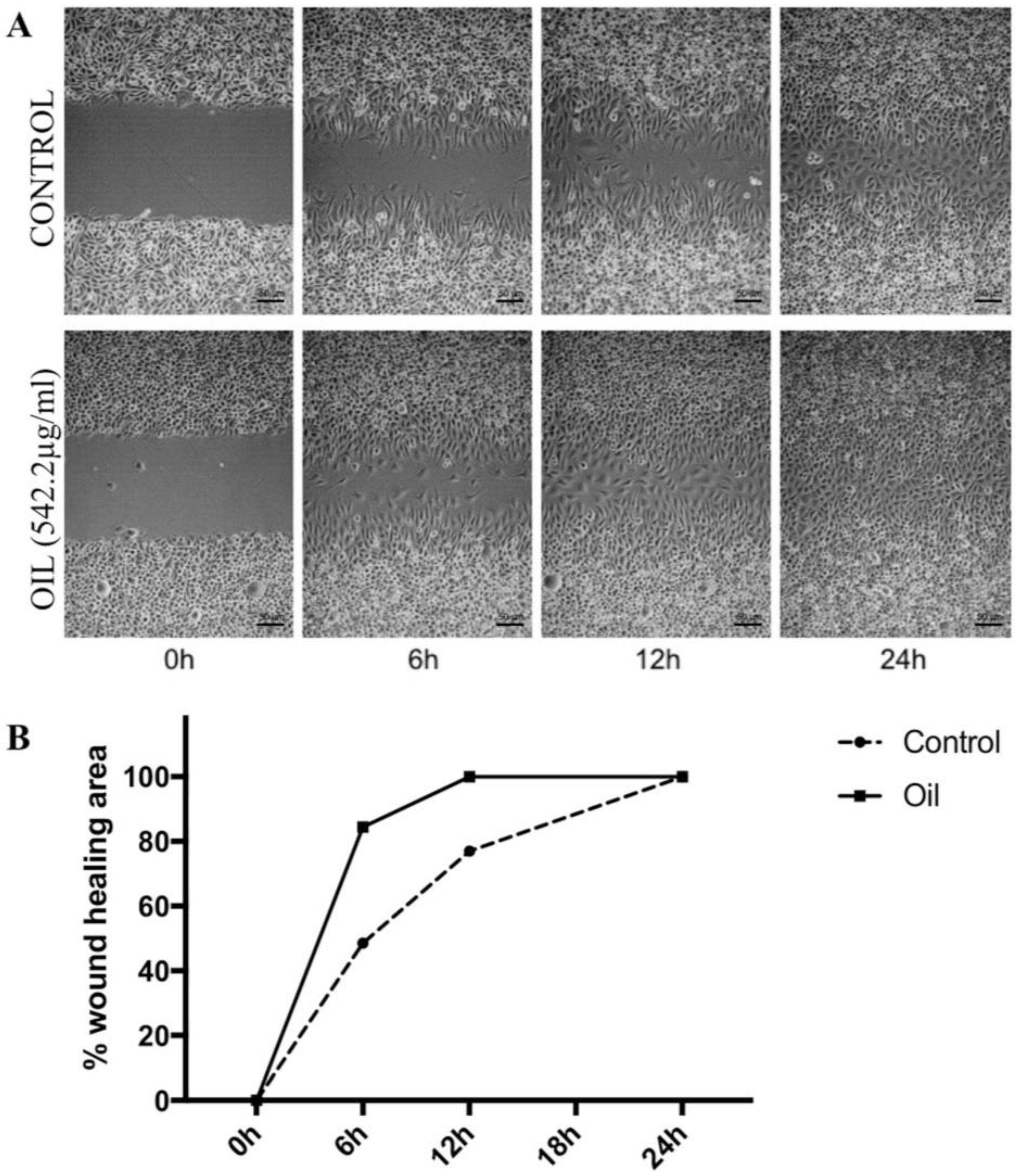
 Molecules Free Full Text Wound Healing Effect Of Essential Oil Extracted From Eugenia Dysenterica Dc Myrtaceae Leaves Html From mdpi.com
The automated image capture. The scratch-wound assay is a simple reproducible assay commonly used to measure basic cell migration parameters such as speed persistence and polarity. Wash the cells with PBS 3 times add SFM 2 ml well incubate in 37oC over night. The scratch assay has been widely used where the closure of the scratch is followed for a set period. An artificial gap is generated on a confluent cell monolayer and movement tracked via microscopy or other imaging. Scratch wound assay creates a gap in confluent monolayer of keratinocytes to mimic a wound.
In this protocol Steps 19 describe the basic method of the in vitro scratch assay for measuring migration of cell populations.
The automated image capture. The automated image capture. There are numerous methods for performing these assays including scratch assays performed on a monolayer of cells adhered to plasticware microplates or culture inserts or using 3D cell culture models. Live cell imaging on migrating cells is also possible using the scratch migration assay. In vitro scratch assay The in vitro scratch assay is a straightforward and economical method to study cell migration in vitro. Cell culture preparation scratch wound assay data acquisition and data analysis.
 Source: biocat.com
This method is based on the observation that upon creation of a new artificial gap so called scratch on a confluent cell monolayer the cells on the edge of. Seed the cells in 8 well plate Nunc 174934 culture until confluent. Fast and easy optimization of culture conditions in scratch assays allows researchers to save time and effort in the protocol optimization phase of their research. This method is based on the observation that upon creation of a new artificial gap so called scratch on a confluent cell monolayer the cells on the edge of. In this protocol Steps 19 describe the basic method of the in vitro scratch assay for measuring migration of cell populations.
 Source: trends.medicalexpo.com
In this protocol Steps 19 describe the basic method of the in vitro scratch assay for measuring migration of cell populations. Scratch wounds heal when cells on either side of the wound migrate into the empty space. Scratch Wound Assay General Protocol Stack Lab 1. This method is based on the observation that upon creation of a new artificial gap so called scratch on a confluent cell monolayer the cells on the edge of. Using the CellComb scratch assay monolayers of NIH3T3 cells were scratchedwounded in a one-direction left or two-direction right pattern.
 Source: sciencedirect.com
Over time migrating cells will fill in the scratched sections and quantified using simple cell counting. Pretreat the cells if desired. Scratch Wound Assay General Protocol Stack Lab 1. It can do this under the influence of a chemoattractant gradient and secretion of proteases. In this proof-of-concept study the effect of different culture conditions on fibroblast scratch closure was investigated.
 Source: researchgate.net
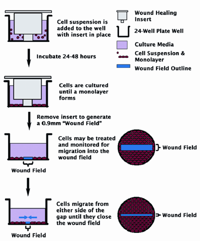
In this protocol Steps 19 describe the basic method of the in vitro scratch assay for measuring migration of cell populations. U2OS cells were seeded in 24-well plates at 2 10 5 cellsmL and incubated for 2448 h. Fast and easy optimization of culture conditions in scratch assays allows researchers to save time and effort in the protocol optimization phase of their research. The optical microscope enables dependable and cheap detection of cell migration. Cells are grown to confluence and a thin wound introduced by scratching with a pipette tip.
 Source: biotekchina.com.cn
Scratch Assay protocol Based on Scratch Wound Healing Assay Martinotti Ranzato Supplies. Over time migrating cells will fill in the scratched sections and quantified using simple cell counting. In Steps 1013 the method is adopted to track migration of indivi- dual cells at the leading edge of the scratch. The scratch assay has been a staple method developed for the purposes of studying cell migration and proliferation patterns in vitro. Using the CellComb scratch assay monolayers of NIH3T3 cells were scratchedwounded in a one-direction left or two-direction right pattern.
 Source: mdpi.com
This assay is an excellent method for characterizing and comparing the movement of cells under various controlled parameters. U2OS cells were seeded in 24-well plates at 2 10 5 cellsmL and incubated for 2448 h. An artificial gap is generated on a confluent cell monolayer and movement tracked via microscopy or other imaging. In this protocol Steps 19 describe the basic method of the in vitro scratch assay for measuring migration of cell populations. The scratch assay has been a staple method developed for the purposes of studying cell migration and proliferation patterns in vitro.
 Source: mdpi.com
The automated image capture. Here we describe a procedure to test the cell migration in vitro with an automated optical camera microscope. Using the CellComb scratch assay monolayers of NIH3T3 cells were scratchedwounded in a one-direction left or two-direction right pattern. In this protocol Steps 19 describe the basic method of the in vitro scratch assay for measuring migration of cell populations. Scratch wound assay creates a gap in confluent monolayer of keratinocytes to mimic a wound.
 Source: biotekchina.com.cn
Scratch wound assay creates a gap in confluent monolayer of keratinocytes to mimic a wound. Cells at the wound edge polarise and migrate into the wound space. The in vitro scratch assay is an easy low-cost and well-developed method to measure cell migration in vitro. Cell culture preparation scratch wound assay data acquisition and data analysis. Scratch Wound Assay General Protocol Stack Lab 1.
 Source: researchgate.net
Cells are grown to confluence and a thin wound introduced by scratching with a pipette tip. The protocol of scratch wound is based on few steps. The automated image capture. In Steps 1013 the method is adopted to track migration of. Using the CellComb scratch assay monolayers of NIH3T3 cells were scratchedwounded in a one-direction left or two-direction right pattern.
 Source: abcam.com
The wound healing or scratch assay is a method to measure two-dimensional cell migration. Wash the cells with PBS 3 times add SFM 2 ml well incubate in 37oC over night. In vitro scratch assay The in vitro scratch assay is a straightforward and economical method to study cell migration in vitro. The automated image capture. Scratch wound assay creates a gap in confluent monolayer of keratinocytes to mimic a wound.
 Source: creative-bioarray.com
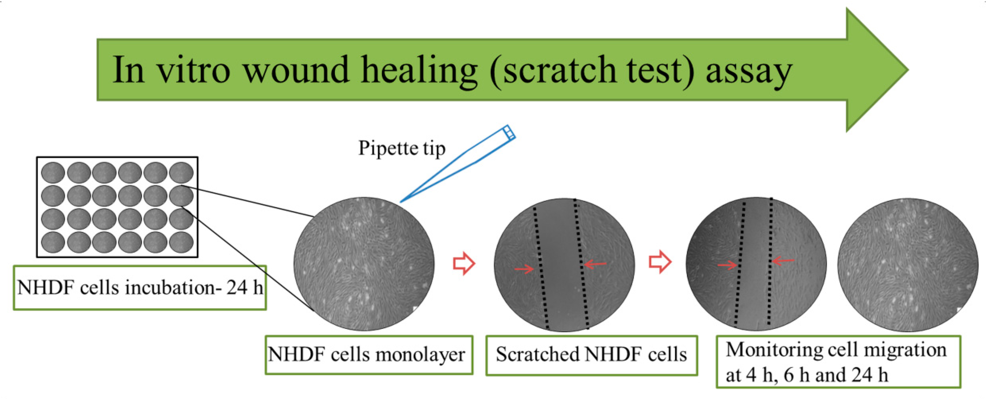
12-Well culture plates TrypsinEDTA Trypsin neutralizing solution HEPES-BSS or PBS 15 or 50 mL conical tube Fresh media 1-2 mL per each well of a 12-well culture plate. The basic steps involve creating a scratch in a cell monolayer capturing the images at the beginning and at regular intervals during cell migration to close the scratch and comparing the images to quantify the migration rate of the cells. Scratch wound assay creates a gap in confluent monolayer of keratinocytes to mimic a wound. Scratch wounds heal when cells on either side of the wound migrate into the empty space. In Steps 1013 the method is adopted to track migration of indivi- dual cells at the leading edge of the scratch.
 Source: europepmc.org
Pretreat the cells if desired. The protocol of scratch wound is based on few steps. Seed the cells in 8 well plate Nunc 174934 culture until confluent. Wash the cells with PBS 3 times add SFM 2 ml well incubate in 37oC over night. Scratch Wound Assay General Protocol Stack Lab 1.
 Source: researchgate.net
Scratch wound assay creates a gap in confluent monolayer of keratinocytes to mimic a wound. In this protocol Steps 19 describe the basic method of the in vitro scratch assay for measuring migration of cell populations. The protocol of scratch wound is based on few steps. Scratch Assay protocol Based on Scratch Wound Healing Assay Martinotti Ranzato Supplies. The basic steps involve creating a scratch in a cell monolayer capturing the images at the beginning and at regular intervals during cell migration to close the scratch and comparing the images to quantify the migration rate of the cells.
Source:
The automated image capture. The scratch-wound assay is a simple reproducible assay commonly used to measure basic cell migration parameters such as speed persistence and polarity. The automated image capture. There are numerous methods for performing these assays including scratch assays performed on a monolayer of cells adhered to plasticware microplates or culture inserts or using 3D cell culture models. In this protocol Steps 19 describe the basic method of the in vitro scratch assay for measuring migration of cell populations.
 Source: researchgate.net
12-Well culture plates TrypsinEDTA Trypsin neutralizing solution HEPES-BSS or PBS 15 or 50 mL conical tube Fresh media 1-2 mL per each well of a 12-well culture plate. An artificial gap is generated on a confluent cell monolayer and movement tracked via microscopy or other imaging. The automated image capture. The scratch assay has been widely used where the closure of the scratch is followed for a set period. Live cell imaging on migrating cells is also possible using the scratch migration assay.
 Source: sciencedirect.com
Scratch wound assay creates a gap in confluent monolayer of keratinocytes to mimic a wound. The wound healing or scratch assay is a method to measure two-dimensional cell migration. Scratch wounds heal when cells on either side of the wound migrate into the empty space. Scratch Wound Assay General Protocol Stack Lab 1. In this protocol Steps 19 describe the basic method of the in vitro scratch assay for measuring migration of cell populations.
 Source: biotekchina.com.cn
In Steps 1013 the method is adopted to track migration of indivi- dual cells at the leading edge of the scratch. Scratch Assay protocol Based on Scratch Wound Healing Assay Martinotti Ranzato Supplies. In Steps 1013 the method is adopted to track migration of indivi- dual cells at the leading edge of the scratch. Fast and easy optimization of culture conditions in scratch assays allows researchers to save time and effort in the protocol optimization phase of their research. In vitro scratch assay The in vitro scratch assay is a straightforward and economical method to study cell migration in vitro.
 Source: ibidi.com
In this proof-of-concept study the effect of different culture conditions on fibroblast scratch closure was investigated. The scratch assay has been widely used where the closure of the scratch is followed for a set period. Seed the cells in 8 well plate Nunc 174934 culture until confluent. The scratch-wound migration assay is based on the formation of a scratch wound across a monolayer of cells and the assessment of how long it takes for the scratch wound to heal. The optical microscope enables dependable and cheap detection of cell migration.
This site is an open community for users to do submittion their favorite wallpapers on the internet, all images or pictures in this website are for personal wallpaper use only, it is stricly prohibited to use this wallpaper for commercial purposes, if you are the author and find this image is shared without your permission, please kindly raise a DMCA report to Us.
If you find this site helpful, please support us by sharing this posts to your preference social media accounts like Facebook, Instagram and so on or you can also bookmark this blog page with the title scratch assay protocol by using Ctrl + D for devices a laptop with a Windows operating system or Command + D for laptops with an Apple operating system. If you use a smartphone, you can also use the drawer menu of the browser you are using. Whether it’s a Windows, Mac, iOS or Android operating system, you will still be able to bookmark this website.


