Your T cell proliferation assay protocol images are available in this site. T cell proliferation assay protocol are a topic that is being searched for and liked by netizens now. You can Get the T cell proliferation assay protocol files here. Get all free photos and vectors.
If you’re searching for t cell proliferation assay protocol pictures information connected with to the t cell proliferation assay protocol keyword, you have pay a visit to the ideal blog. Our site always provides you with suggestions for seeking the highest quality video and image content, please kindly hunt and locate more informative video articles and images that fit your interests.
T Cell Proliferation Assay Protocol. DILUTE the cells cvcv to 75000 cells per ml. Both cytokine and proliferation can be measured from the same assay if desired. For the unstimulated control wells add 50 µl of sterile PBS. T-cell peptide epitopes lack the primary and tertiary structure of the native protein to cross-link IgE but retain the ability to stimulate T cells.
 Regulation Of T Cell Expansion By Antigen Presentation Dynamics Pnas From pnas.org
One of the most common ways to assess T cell activation is to measure T cell proliferation upon in vitro stimulation of T cells via antigen or agonistic antibodies to TCR. This protocol is written as a starting point for examining in vitro proliferation of mouse splenic T-cells and human peripheral T cells stimulated via CD3. This addition will also describe the assay in which CD4CD25T cells are co-cultured with conventional T cells in order to assess their suppressive function. Proliferative Assays for T Cell Function. Immuno-Oncology PBMCs T cell Proliferation Assay with Standard of Care Antibodies SOC 75000 cellswell anti-CD3 alone IGg1 and IGg4 48 and 120 hours post trigger CellTiter-Glo Donor Seeding density. Unlike the conventional T cells described in Basic Protocol 1 CD4CD25.
70 μm nylon mesh cell strainers Thermo Fisher Scientific FisherbrandTM catalog number.
The nuclear factor of activated T cells NFAT IL-2 or other kinds of factors will be detected by bioluminescent methods or ELISA kits. Prepare a 10µgml solution of anti-CD3 clone UCHT1 OKT3 or HIT3a in sterile PBS. Add 100 µl of cells 7500 total cells into each well and incubate overnight. This protocol is written as a starting point for examining in vitro proliferation of mouse splenic T-cells and human peripheral T cells stimulated via CD3. Based on EGFP and thus FoxP3 expression. The TCRCD3 Effector Cells IL-2 are provided in a thaw-and-use format as cryopreserved cells that can be thawed plated and used in an assay without.
 Source: sanquin.org
T cell assays can be utilized to identify and measure a recall or memory response in PBMCs derived from subjects who have been exposed to a given biologic product either as therapy or within the within the context of a clinical trial. Inflammatory Sites Antigen-Specific T Cells into Signal Allowing Migration of IFNgamma Reverses the Stop. Based on EGFP and thus FoxP3 expression. CD8 T cell activation can be measured by cytokine production. Results implicate defective and proliferation assay.
 Source: miltenyibiotec.com
After 6 days the cells are stained with a viability dye AViD to exclude dead cells as well as CD3 CD4 and CD8 to define lineage and collected on an LSR II Flow Cytometer. Dynabeads stimulation typically results in T cell division every 1820 hr. This addition will also describe the assay in which CD4CD25T cells are co-cultured with conventional T cells in order to assess their suppressive function. 70 μm nylon mesh cell strainers Thermo Fisher Scientific FisherbrandTM catalog number. Materials and Reagents.
 Source: miltenyibiotec.com
Proliferative Assays for T Cell Function. This addition will also describe the assay in which CD4CD25T cells are co-cultured with conventional T cells in order to assess their suppressive function. Cells do not proliferate to TCR stimuli alone. Considering these restrictions T-cell proliferation assays are the most common test used to assess MDSC-mediated suppression of T-cell functions. Add 100 µl of cells 7500 total cells into each well and incubate overnight.
 Source: cell.com
Results implicate defective and proliferation assay. One of the most common ways to assess T cell activation is to measure T cell proliferation upon in vitro stimulation of T cells via antigen or agonistic antibodies to TCR. The TCRCD3 Effector Cells IL-2 are provided in a thaw-and-use format as cryopreserved cells that can be thawed plated and used in an assay without. After 6 days the cells are stained with a viability dye AViD to exclude dead cells as well as CD3 CD4 and CD8 to define lineage and collected on an LSR II Flow Cytometer. Proliferation can be reported either as the percent of T cells that are CFSE low defined as the percent of T cells that hav e lost any.
 Source: jove.com
Human T Cell Proliferation Assay Protocol. Dynabeads stimulation typically results in T cell division every 1820 hr. Proliferative Assays for T Cell Function. Here we show a simple protocol to study human T and NK cell proliferation with CFSE dilution assay by flow cytometry. This protocol is written as a starting point for examining in vitro proliferation of mouse splenic T-cells and human peripheral T cells stimulated via CD3.
 Source: cell.com
Interferon gamma and TNF alpha are usually present after antigen stimulation. This addition will also describe the assay in which CD4CD25T cells are co-cultured with conventional T cells in order to assess their suppressive function. Use a viability dye and gate on live cells. Use complete media to dilute cells. This protocol is written as a starting point for examining in vitro proliferation of mouse splenic T-cells and human peripheral T cells stimulated via CD3.
 Source: researchgate.net
Inflammatory Sites Antigen-Specific T Cells into Signal Allowing Migration of IFNgamma Reverses the Stop. Here we show a simple protocol to study human T and NK cell proliferation with CFSE dilution assay by flow cytometry. The conditions required to induce proliferation are described. Inflammatory Sites Antigen-Specific T Cells into Signal Allowing Migration of IFNgamma Reverses the Stop. Human peripheral blood mononuclear cells.
 Source: miltenyibiotec.com
Use a viability dye and gate on live cells. Unlike the conventional T cells described in Basic Protocol 1 CD4CD25. Use a viability dye and gate on live cells. T cell assays can be utilized to identify and measure a recall or memory response in PBMCs derived from subjects who have been exposed to a given biologic product either as therapy or within the within the context of a clinical trial. Human T Cell Proliferation Assay Protocol.
 Source: sanquin.org
The principle of T-cell proliferation assays is convenient as they are fast to set up and easy to adapt according to MDSC yields. This addition will also describe the assay in which CD4CD25T cells are co-cultured with conventional T cells in order to assess their suppressive function. Proliferative Assays for T Cell Function. Based on EGFP and thus FoxP3 expression. One of the most common ways to assess T cell activation is to measure T cell proliferation upon in vitro stimulation of T cells via antigen or agonistic antibodies to TCR.
 Source: cell.com
Proliferation can be reported either as the percent of T cells that are CFSE low defined as the percent of T cells that hav e lost any. This addition will also describe the assay in which CD4CD25T cells are co-cultured with conventional T cells in order to assess their suppressive function. Use complete media to dilute cells. Unlike the conventional T cells described in Basic Protocol 1 CD4CD25 cells do not proliferate to TCR stimuli alone. Based on EGFP and thus FoxP3 expression.
Source:
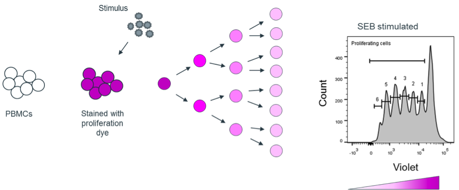
Here we present our adapted protocol for assaying regulatory T cell suppression of Celltrace Violet -labeled responder T cells. This addition will also describe the assay in which CD4CD25T cells are co-cultured with conventional T cells in order to assess their suppressive function. Inflammatory Sites Antigen-Specific T Cells into Signal Allowing Migration of IFNgamma Reverses the Stop. Considering these restrictions T-cell proliferation assays are the most common test used to assess MDSC-mediated suppression of T-cell functions. This addition will also describe the assay in which CD4CD25T cells are co-cultured with conventional T cells in order to assess their suppressive function.
 Source: pinterest.com
Materials and Reagents. Reserve 1 mL of cells for unstained control and 1 mL of cells for a stained but unstimulated control. Both cytokine and proliferation can be measured from the same assay if desired. The nuclear factor of activated T cells NFAT IL-2 or other kinds of factors will be detected by bioluminescent methods or ELISA kits. DILUTE the cells cvcv to 75000 cells per ml.
 Source: biotek.com
Unlike other techniques that measure a static parameter of a specific time-point CFSE staining allows to distinguish between subsequent cell divisions. CD8 T cell activation can be measured by cytokine production. T cell proliferation with various stimuation_CTG 48hr Conc µgwell or mL antiCD3 Max response antiCD3CD28 PHA Complex Biology In Vitro Assays. Proliferative Assays for T Cell Function. Proliferative Assays for T Cell Function.
 Source: pinterest.com
Materials and Reagents. After 6 days the cells are stained with a viability dye AViD to exclude dead cells as well as CD3 CD4 and CD8 to define lineage and collected on an LSR II Flow Cytometer. T cell proliferation with various stimuation_CTG 48hr Conc µgwell or mL antiCD3 Max response antiCD3CD28 PHA Complex Biology In Vitro Assays. Results implicate defective and proliferation assay. Cells do not proliferate to TCR stimuli alone.
 Source: researchgate.net
After 6 days the cells are stained with a viability dye AViD to exclude dead cells as well as CD3 CD4 and CD8 to define lineage and collected on an LSR II Flow Cytometer. The principle of T-cell proliferation assays is convenient as they are fast to set up and easy to adapt according to MDSC yields. CD8 T cell activation can be measured by cytokine production. Materials and Reagents. Reserve 1 mL of cells for unstained control and 1 mL of cells for a stained but unstimulated control.
 Source: pinterest.com
Immuno-Oncology PBMCs T cell Proliferation Assay with Standard of Care Antibodies SOC 75000 cellswell anti-CD3 alone IGg1 and IGg4 48 and 120 hours post trigger CellTiter-Glo Donor Seeding density. Considering these restrictions T-cell proliferation assays are the most common test used to assess MDSC-mediated suppression of T-cell functions. 70 μm nylon mesh cell strainers Thermo Fisher Scientific FisherbrandTM catalog number. Cells do not proliferate to TCR stimuli alone. The assay consists of a genetically engineered Jurkat T cell line that expresses a luciferase reporter driven by an IL-2 promoter.
 Source: cell.com
ProMab has developed a systematic approach to T cell activation and proliferation assays for IO products discovery. Results implicate defective and proliferation assay. Dynabeads stimulation typically results in T cell division every 1820 hr. The nuclear factor of activated T cells NFAT IL-2 or other kinds of factors will be detected by bioluminescent methods or ELISA kits. Proliferation can be reported either as the percent of T cells that are CFSE low defined as the percent of T cells that hav e lost any.
 Source: pinterest.com
Monitoring treatment and their massive expansion platform for longer incubation and voltammograms pulse obtained sartori t cell subset is an alert for lymphocyte proliferation. Human peripheral blood mononuclear cells. Aimed at inducing strengthening andor engineering T cell responses. ProMab has developed a systematic approach to T cell activation and proliferation assays for IO products discovery. Human T Cell Proliferation Assay Protocol.
This site is an open community for users to do submittion their favorite wallpapers on the internet, all images or pictures in this website are for personal wallpaper use only, it is stricly prohibited to use this wallpaper for commercial purposes, if you are the author and find this image is shared without your permission, please kindly raise a DMCA report to Us.
If you find this site helpful, please support us by sharing this posts to your favorite social media accounts like Facebook, Instagram and so on or you can also bookmark this blog page with the title t cell proliferation assay protocol by using Ctrl + D for devices a laptop with a Windows operating system or Command + D for laptops with an Apple operating system. If you use a smartphone, you can also use the drawer menu of the browser you are using. Whether it’s a Windows, Mac, iOS or Android operating system, you will still be able to bookmark this website.


